IOTM: Imaging Through A Subretinal Macular Hemorrhage with Topcon SS-OCT
Image of the Month (IOTM) is a collection of interesting clinical cases with high quality images for all relevant imaging modalities (ex: color fundus, OCT, OCTA, FAF, FA, En Face, Red-free, choroidal vasculography (CVG), anterior imaging) and other clinical results if relevant (ex: visual field plots). Each case is submitted by an eye care professional using one of Topcon’s industry-leading OCT devices.
Case background
64 year-old female presented with acute vision loss in her right eye.
Diagnosis: Wet Age-Related Macular Degeneration
A
Color photograph of the right eye (Topcon TRC-50DX) demonstrating a large subretinal hemorrhage in the macula.
B & C
Swept-Source OCT (Topcon DRI Triton) demonstrating subretinal fluid and a large area of subretinal hyper-reflective material (SRHM) consistent with the hemorrhage seen on color photography. The underlying RPE and choroid is also visible.
The patient was subsequently treated with anti‑VEGF therapy.
Ophthalmic Photographer: John Golding
Images courtesy of Netan Choudhry M.D. FRCS(C) DABO, Medical Director Vitreous Retina Macula Specialists of Toronto (VRM Toronto)


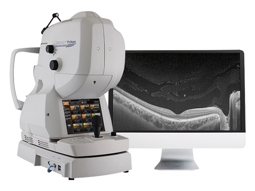
Increase diagnostic capabilities with the multi-modal swept source OCT
OCT Angiography is not available for sale in the US
The opinions, ideas, views and assumptions expressed are the author’s own and do not necessarily represent the views of Topcon, nor do they constitute advice from Topcon.
Recent Posts

IOTM: Choroidal Nevus with Drusen
This 52 year-old patient presented with a sudden central visual…
Read more
IOTM: Branch Retinal Artery Occlusion
This 52 year-old patient presented with a sudden central visual…
Read more
IOTM: Toxoplasmosis Retinochoroiditis
A 57-year-old Indian female with a prior history of posterior…
Read more