IOTM: Toxoplasmosis Retinochoroiditis
Image of the Month (IOTM) is a collection of interesting clinical cases with high quality images for all relevant imaging modalities (ex: color fundus, OCT, OCTA, FAF, FA, En Face, Red-free, choroidal vasculography (CVG), anterior imaging) and other clinical results if relevant (ex: visual field plots). Each case is submitted by an eye care professional using one of Topcon’s industry-leading OCT devices.
Case background
A 57-year-old Indian female with a prior history of posterior uveitis was referred for scarring OD. BCVA 20/20 OU, diagnostic testing demonstrated +IgG antibodies to toxoplasmosis, IgM was negative. Upon further questioning, the patient did report a prior history of eating uncooked pork. Color Fundus photograph of the right eye (Image A) demonstrated several extrafoveal pigmented chorioretinal atrophy lesions. Enface Swept-Source OD (Image B) demonstrated several chorioretinal lesions. OCT B-scan of the macula (Image C) demonstrated outer retinal, ellipsoid zone, RPE, and atrophy of the choriocapillaris. 24mm posterior pole structural B-scan OCT (Image D) revealed two regions of retinal atrophy.
— Netan Choudhry, MD FRCS(C) FASRS
Diagnosis: Toxoplasmosis Retinochoroiditis
Captured with: Topcon DRI OCT Triton Swept-Source
DRI OCT Triton Images:
A. Color Fundus Image
B. Enface Swept-Source OCT Image
C. OCT B-Scan of the Macula
D. OCT B-Scan


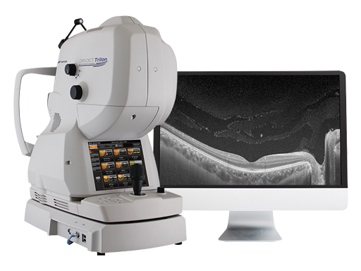
Increase diagnostic capabilities with the multi-modal swept source OCT
The opinions, ideas, views and assumptions expressed are the author’s own and do not necessarily represent the views of Topcon, nor do they constitute advice from Topcon.
Recent Posts

IOTM: Choroidal Nevus with Drusen
This 52 year-old patient presented with a sudden central visual…
Read more
IOTM: Branch Retinal Artery Occlusion
This 52 year-old patient presented with a sudden central visual…
Read more
IOTM: Toxoplasmosis Retinochoroiditis
A 57-year-old Indian female with a prior history of posterior…
Read more