IOTM: Exophytic Papillary Angioma
Image of the Month (IOTM) is a collection of interesting clinical cases with high quality images for all relevant imaging modalities (ex: color fundus, OCT, OCTA, FAF, FA, En Face, Red-free, choroidal vasculography (CVG), anterior imaging) and other clinical results if relevant (ex: visual field plots). Each case is submitted by an eye care professional using one of Topcon’s industry-leading OCT devices.
Case background
A 50-year-old female patient with known history of von Hippel-Lindau disease. Her left eye is blind and the right eye was treated with focal laser coagulation in 1999 for a small retinal angioma in the temporal periphery. Since then no treatment of the right eye was necessary. For some weeks, she has blurred vision and on SD-OCT the general ophthalmologist found some intraretinal fluid next to the optic disc.
The vision was still 20/20.
Diagnosis: Exophytic Papillary Angioma
We performed a 9 x 9mm Swept Source OCT-A(1) using the central fixation to get both macula and optic disc in one scan visualized. The SS-OCT-A of the superficial capillaries (A) was normal but in the deeper layer (B), a vascular structure was detected at the temporal margin of the optic disc. On B-Scan a solid intraretinal mass with confined vascular flow (C, arrow) was shown. There was no contact to RPE and the SS-OCT-A of the sub-RPE layers was normal, so that a juxtapapillary CNV could be excluded. The diagnosis of an exophytic papillary angioma was made. The dimension of the angioma (BxLxH 640 x 1028 x 276μm) was measured by vertical and horizontal B-Scans (D, E). The en-face mode (F) demonstrated much more detailed information about the two-dimensional extend of the angioma and the surrounding exudation compared to the fundus photo (G).
Because of the symptomatic clinical presentation, a systemic treatment with propranolol hydrochloride (3mg/kg) was initiated.
— Ass. Prof. Gerasimos Anastassiou, MD, PhD


OCT Angiography is not available for sale in the US
The opinions, ideas, views and assumptions expressed are the author’s own and do not necessarily represent the views of Topcon, nor do they constitute advice from Topcon.
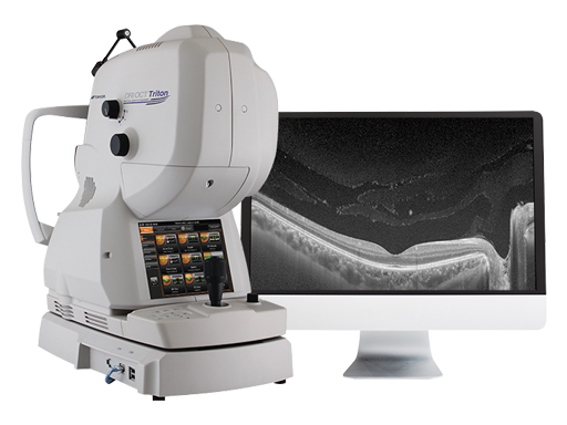
Increase diagnostic capabilities with the multi-modal swept source OCT
Recent Posts
IOTM: Choroidal Nevus with Drusen
This 52 year-old patient presented with a sudden central visual…
Read more
IOTM: Branch Retinal Artery Occlusion
This 52 year-old patient presented with a sudden central visual…
Read more
IOTM: Toxoplasmosis Retinochoroiditis
A 57-year-old Indian female with a prior history of posterior…
Read more