IOTM: Central Serous Retinopathy Secondary to Eczema Treatment
Image of the Month (IOTM) is a collection of interesting clinical cases with high quality images for all relevant imaging modalities (ex: color fundus, OCT, OCTA, FAF, FA, En Face, Red-free, choroidal vasculography (CVG), anterior imaging) and other clinical results if relevant (ex: visual field plots). Each case is submitted by an eye care professional using one of Topcon’s industry-leading OCT devices.
Case background
A 45-year-old male currently taking topical prednisone cream for eczema was referred to our clinic for distorted and blurry vision in his right eye, testing at 20/80 OD.
Swept Source OCT from the Topcon DRI OCT Triton (Images D & E) demonstrated a thickened choroid and subretinal fluid as well as subretinal hyperreflective material overlying the fovea.
Fundus autofluorescence imaging (Image A) showed central hyperautofluorescence consistent with subretinal fluid, confirming the diagnosis of Central Serous Retinopathy.
Diagnosis: Central Serous Retinopathy
Captured with: Topcon DRI OCT Triton & TRC-50DX
Image Glossary
A. Fundus Autofluorescence Image (TRC-50DX)
B. OCT EnFace Image (Triton)
C. 50O True Color Fundus Photo (TRC- 50DX)
D. Swept Source OCT Horizontal B-Scan (Triton)
E. Swept Source OCT Vertical B-Scan (Triton)
Images courtesy of Netan Choudhry M.D. FRCS(C) DABO, Medical Director Vitreous Retina Macula Specialists of Toronto (VRM Toronto)


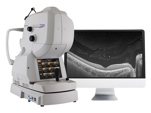
Increase diagnostic capabilities with the multi-modal swept source OCT
Recent Posts

IOTM: Choroidal Nevus with Drusen
This 52 year-old patient presented with a sudden central visual…
Read more
IOTM: Branch Retinal Artery Occlusion
This 52 year-old patient presented with a sudden central visual…
Read more
IOTM: Toxoplasmosis Retinochoroiditis
A 57-year-old Indian female with a prior history of posterior…
Read more