Case Study – Advanced PDR with Non-Perfusion and Macular Edema
Topcon Sponsored
Background
33-year-old Caucasian female with history of myopia, vulvar cancer, family history of diabetes and liver cancer presented with a visual acuity of 20/200 (OD), 20/50 (OS). Intraocular pressures were measured 17mmHg (OD), 19mmHg (OS). A posterior subcapsular cataract was observed with all other ocular physiological characteristics normal.
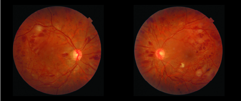
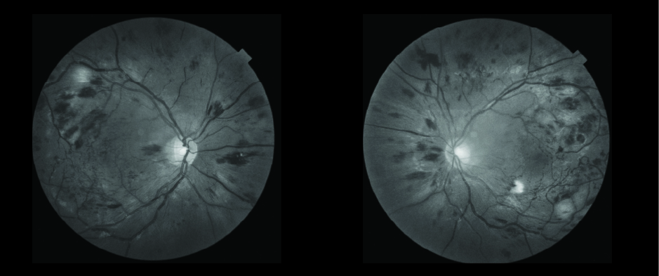
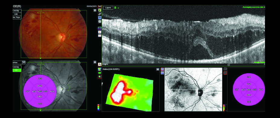
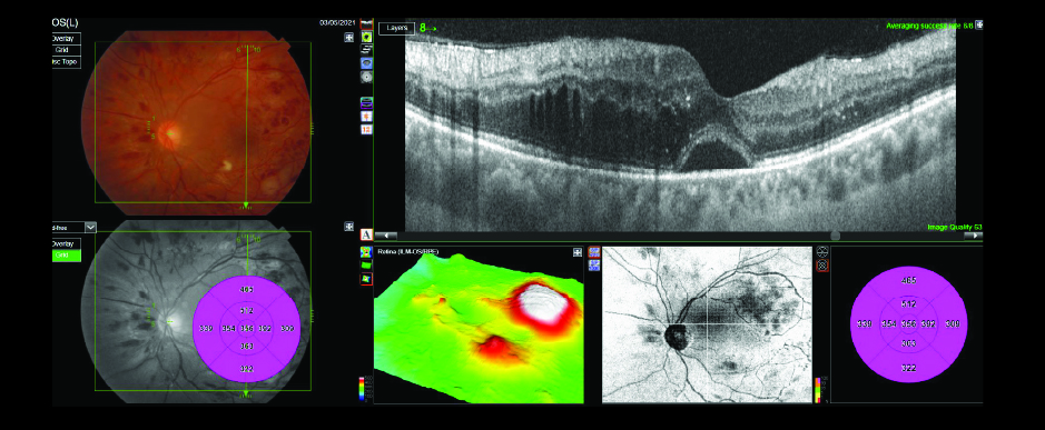
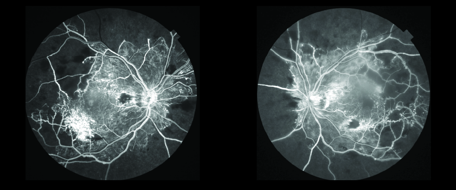
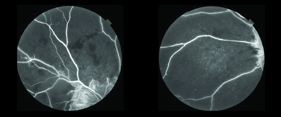
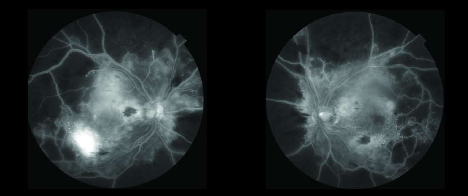
3D Wide OCT Combination Scans, fluorescein angiography and fundus photos were obtained OU. Macular edema, subretinal fluid, intraretinal hemorrhages, cotton wool spots (CWS), nonperfusionable vessels and hemorrhages in the peripheral vessels were visualized OU.
Diagnosis: Advanced Proliferative Diabetic Retinopathy (PDR), retinal non-perfusion and macular edema OU
Treatment: Bevacizumab, PRP laser (OU)
Discussion: 45-degree fluorescein images, true-color photos of the retina and penetrating swept-source OCT scans helped to confirm and document the clinical findings
Captured with
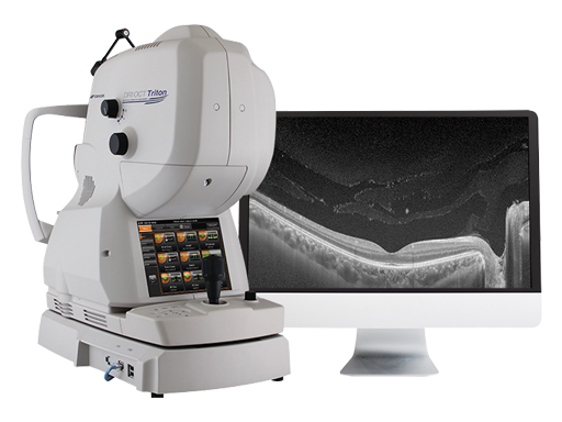
Topcon DRI Triton Swept-Source OCT Fundus Camera (October 2022)
Case and Images Courtesy of
Jonathan Barofsky, MD, FACS
Retina Care Center Lakewood, NJ USA
*The opinions, ideas, views and assumptions expressed are the author’s own and do not
necessarily represent the views of Topcon, nor do they constitute advice from Topcon.
Increase diagnostic capabilities with the multi-modal swept source OCT
Recent Posts

VIDEO: How Technology is Advancing Myopia Management in Primary Eye Care
Dr. Jason Compton is dedicated to being on the front…
Read more
Advancing Glaucoma Care: A Collaborative Approach to Data-Driven Decision Support
Michael Chaglasian, OD, Associate Professor at the Illinois College of…
Read more
VIDEO: Why Axial Length Tracking is a Game-Changer for Myopia Management
Discover how Dr. Ashley Tucker elevated her myopia management practice…
Read more