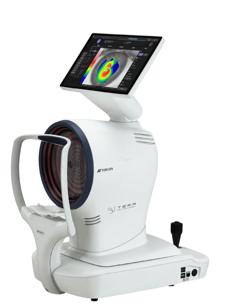
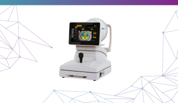
Corneal Analyzer + Dry Eye Diagnostic Suite
Automated to delegate testing. Designed to empower decisions. Optimized to drive treatment acceptance.
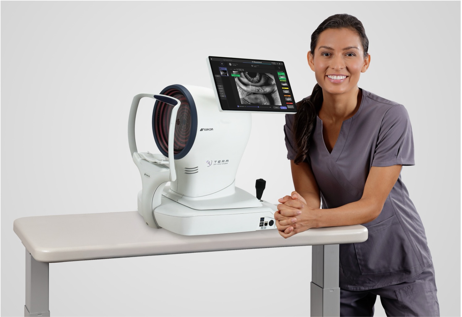
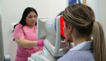
The TERA Dry Eye Imager is a multimodal platform purpose-built to detect, grade, and manage dry eye disease. Powered by robotic automation and high-resolution imaging, TERA standardizes capture, streamlines workflow, and delivers clear, actionable guidance for treatment and follow-up.
Watch this TERA overview
Streamline Workflow with Automation
Automated to delegate testing and save time in the exam lane.
Build Clinical Confidence
Designed to empower confident clinical decisions with data-driven insights following new DEWS III guidelines.¹
Fuel Practice Growth
Optimized to improve treatment acceptance and drive emerging revenue opportunities.
Key Features
- CORNEAL ANALYSIS: Captures corneal topography, anterior corneal wavefront (Zernike) analysis, and white-to-white measurement in a single capture to support diagnosis and monitoring of corneal irregularity and guide custom contact lens fitting.
- DYNAMIC AND STATIC PUPILLOMETRY: Measures pupil size, centration, and light response under various conditions to support diagnosis and patient discussions.
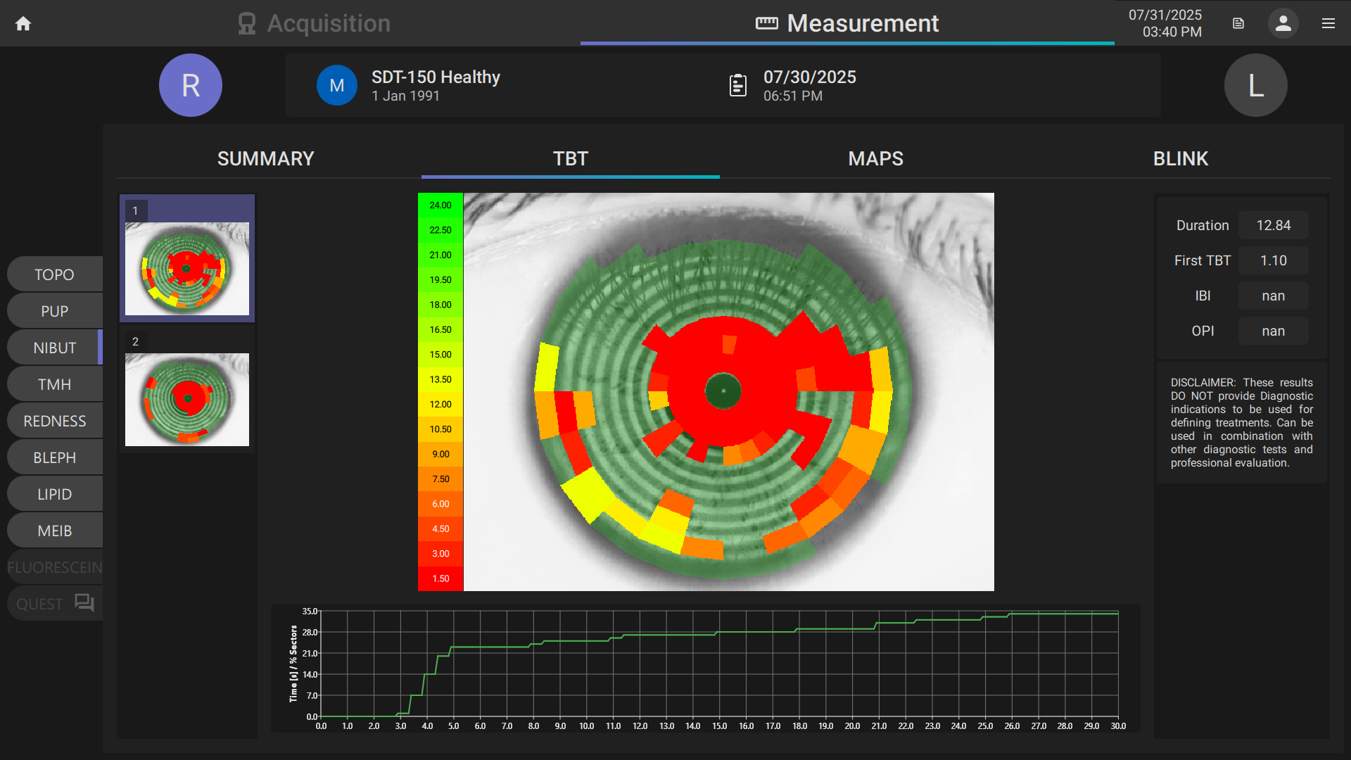
- NON-INVASIVE TEAR BREAK-UP TIME (NIBUT): Automated capture of tear film stability for dry eye screening and diagnosis.
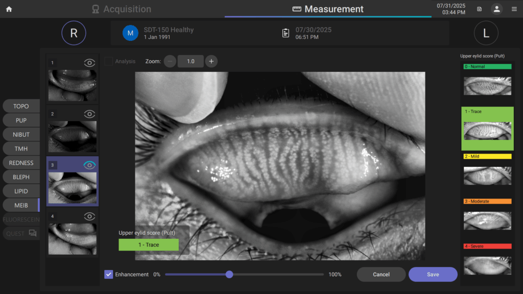
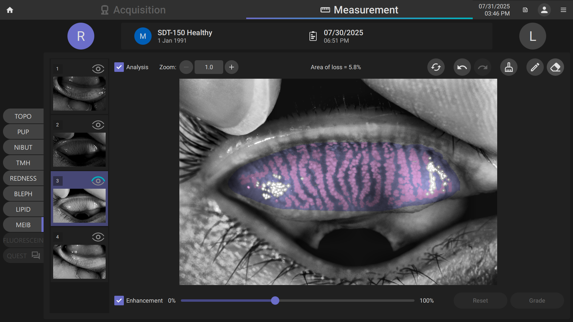
- MEIBOMIAN GLAND IMAGING AND ANALYSIS: Visualizes and evaluates meibomian gland structure and loss.
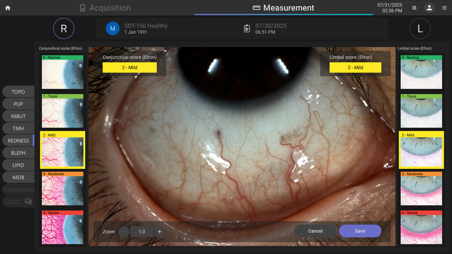
- HIGH-RESOLUTION COLOR IMAGING: Supports evaluation of blepharitis and conjunctival redness to aid diagnosis, documentation, and patient education.
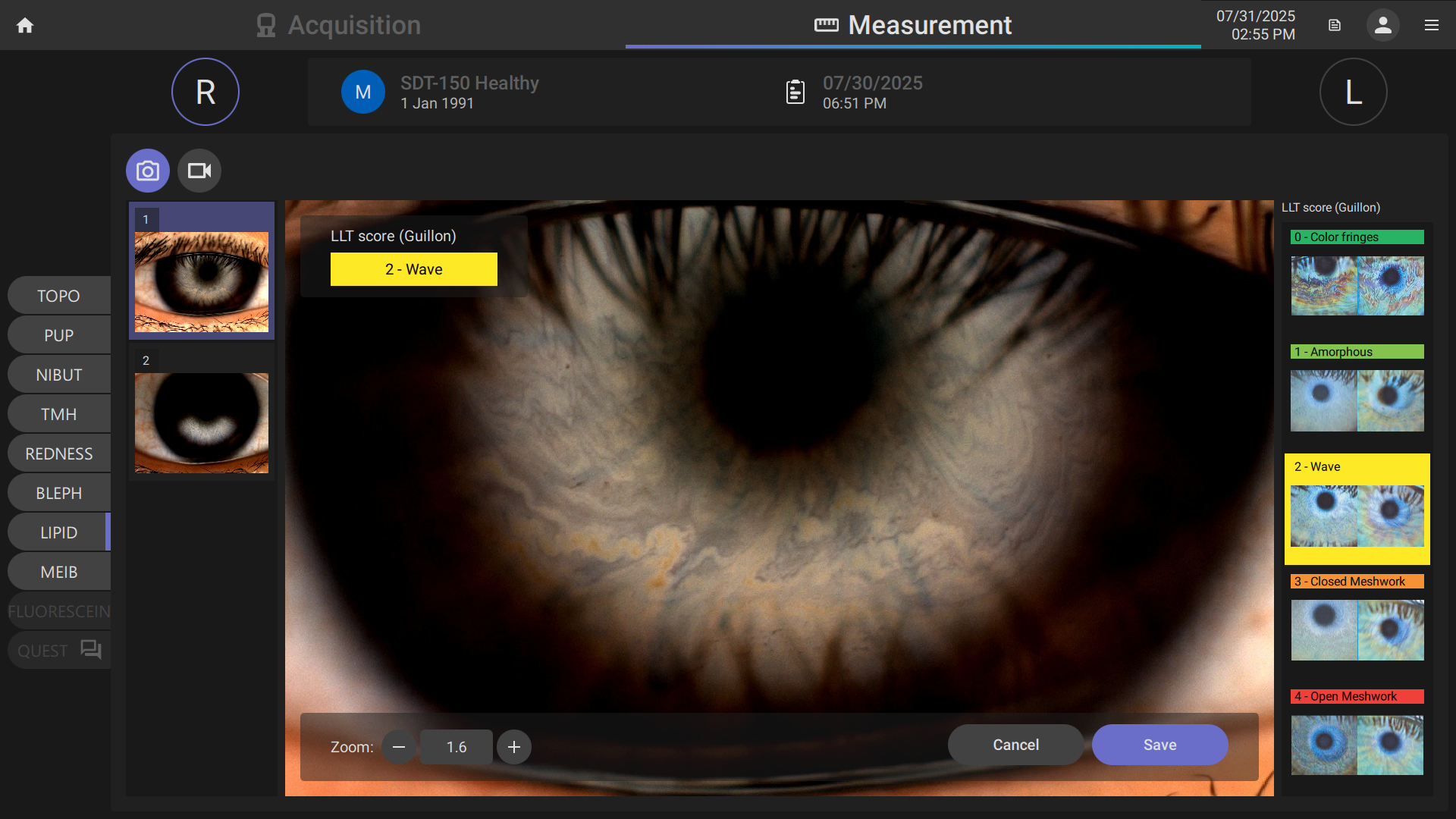
- PATENTED DIFFUSED WHITE LIGHT: Provides uniform illumination that minimizes glare and enhances image clarity, delivering clear visualization of the tear film lipid layer.
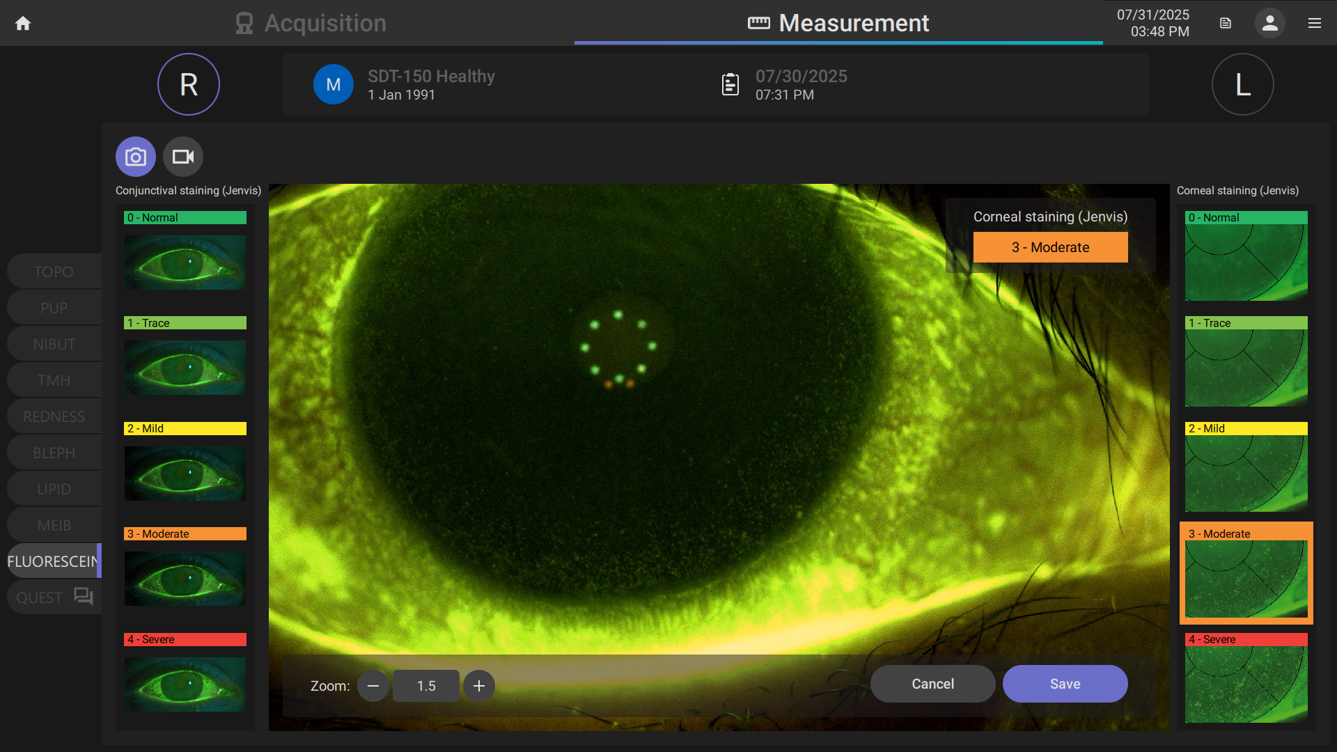
- FLUORESCEIN IMAGING: Evaluates signs of ocular surface staining and enhances assessment of contact lens fit.
- VALIDATED CLINICAL GRADING SCALES: On-screen access to clinically validated scales (Efron, JENVIS, Meiboscale, Guillon) ensures consistent evaluation, documentation, and patient communication.
- INTEGRATED DRY EYE QUESTIONNAIRES (OSDI-6 and DEQ-5): Effectively capture patient-reported symptoms with validated dry eye questionnaires, including the OSDI-6 survey recommended by DEWS III guidelines.
- TEAR MENISCUS HEIGHT: Quantifies tear volume to identify drivers of dry eye disease and assess aqueous insufficiency.
¹ Wolffsohn JS, Benítez-Del-Castillo J, Loya-Garcia D, et al. TFOS DEWS III Diagnostic Methodology. Am J Ophthalmol.
Ready to learn more?

Contact us today
TERA Dry Eye Imager™ Brochure
TERA Dry Eye Imager™ Flyer
Related Articles
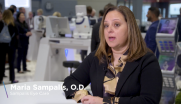
Maria Sampalis, OD shares why the Topcon CA‑800 Corneal Analyzer was a…
Optometrist Celesta Ferreira, owner of Cypress Optique in Cypress, Texas,…
“Building my dry eye practice has drastically impacted my bottom…