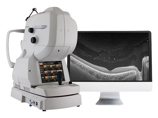
Multimodal Swept-Source OCT
A New Frontier in OCT Imaging
Triton uses Swept-Source technology to allow visualization into the deepest layers of the eye – even through cataracts, hemorrhages, gas bubbles, and other media opacities, making it possible for more patients to be imaged.
The fast 100 kHz scanning speed and invisible scan beam rapidly capture detailed images, resulting in fewer motion artifacts and stunning image quality. Decrease chair time and improve your clinical workflow with fewer rescans and multimodal imaging including, SS-OCT, True-Color, Digital Red-free, and optional Anterior Segment OCT.
Triton (plus) adds fluorescein angiography (FA) and fundus autofluorescence (FAF) to the Triton.
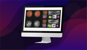
Watch this quick Triton demo
Product Features

Deep penetration through media opacities such as cataracts and hemorrhages

Multimodal Imaging: SS-OCT + Non-mydriatic true color fundus imaging, FA and FAF available²

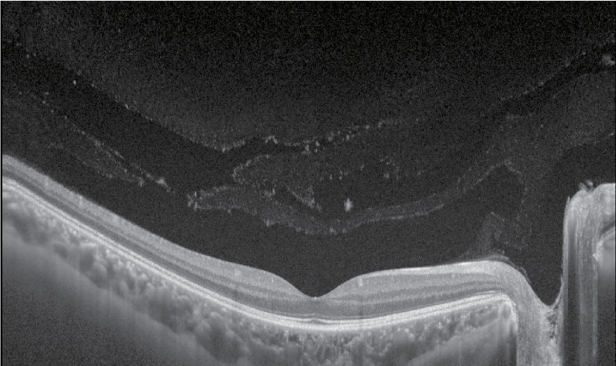
Stunningly detailed images with 100k A-scans/sec. and 1,050nm wavelength

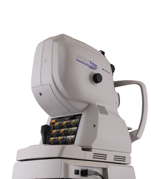
Invisible scan beam allows patients to focus on the fixation target and reduce involuntary eye movement
The Triton in Clinical Practice
Key Features
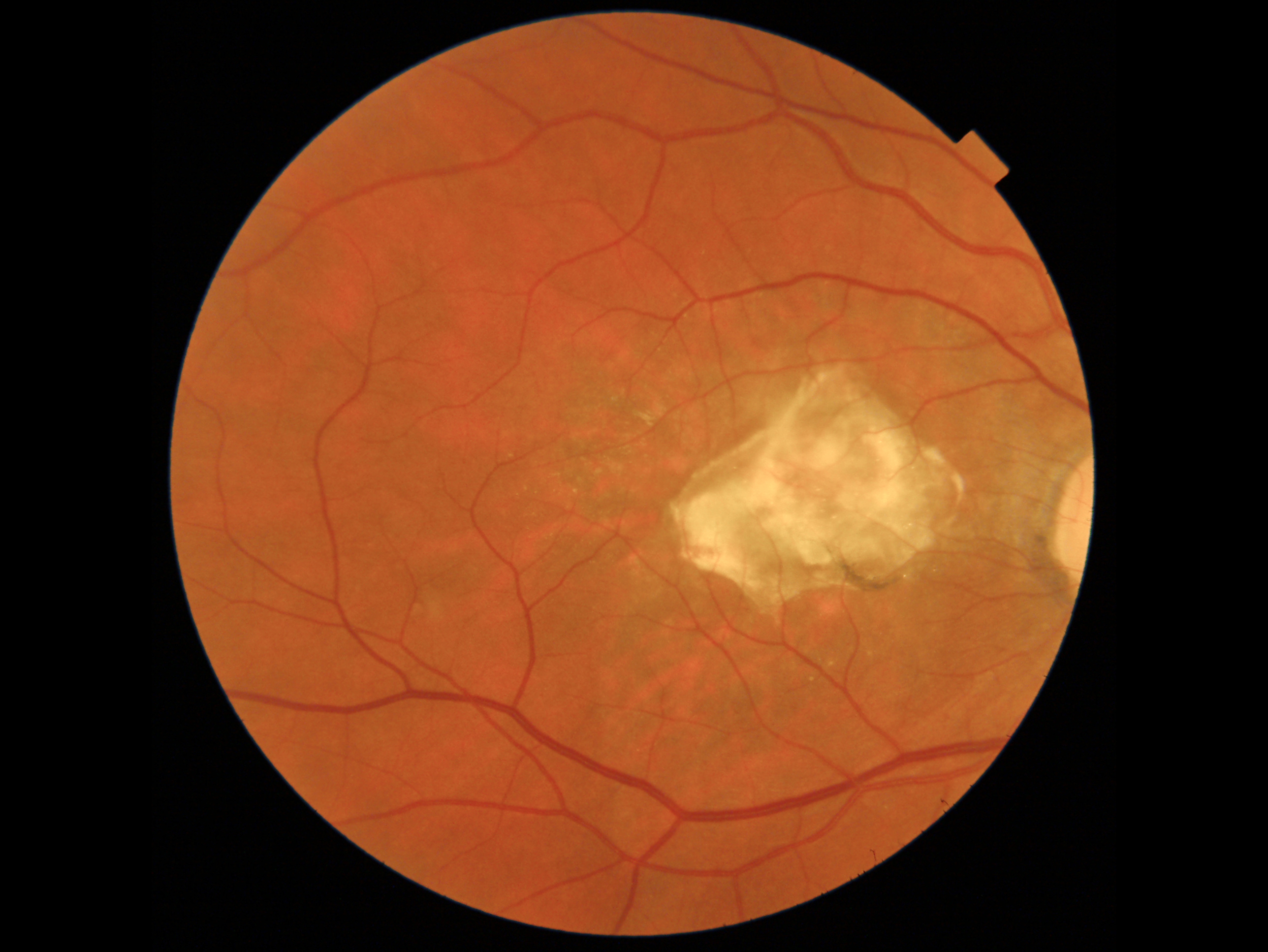
- HIGH RESOLUTION: Multimodal platform provides easy, yet comprehensive comparison of microvascular impairment with FA1, FAF1, OCT and color fundus images.
- SCAN MORE PATIENTS: Swept-source technology allows imaging through media opacities.
- FEWER RESCANS: Invisible scan beam allows patients to focus on the fixation target and reduce involuntary eye movement.
- WIDE SCAN: A 12mm x 9mm scan encompasses the optic nerve and macula and can be acquired in 1.8 seconds for fast assessment of the posterior pole.
- RICH, DETAILED IMAGES: Image quality is further enhanced by PixelSmart® Technology2.
1DRI OCT Triton Plus only.
2PixelSmart® is a function of IMAGEnet 6 software.
Ready to learn more?

Contact us today
| TRITON SPECIFICATIONS | |
Fundus Imaging | |
| Field of View | 45° / 30° (Digital Zoom) |
| Operating Distance | 34.8mm |
| Minimum Pupil Diameter | Ø4.0mm / Small Pupil Mode: Ø3.3mm |
| Resolution (On Fundus) | Center: 60 Lines/mm or more, |
OCT | |
| Scan Range (On Fundus) | 6 to 12mm |
| Scan Patterns | 3D Wide: 12x9mm |
| Scan Speed | 100,000 A-Scans Per Second |
| Lateral Resolution | 20 μm |
| Axial Resolution | Optical: 8 μm |
| Minimum Pupil Diameter | Ø2.5mm |
| Fixation Target | Internal Fixation Target/ Peripheral Fixation Target / External Fixation Target |
| Diopter Range | Without the diopter compensation lens: -13D to +12D |
Anterior Segment | |
| Photography Type | IR |
| Operating Distance | 17 mm |
| Scan Range (On Cornea) | 3 to 16 mm |
| Scan Patterns | Line Anterior Segment: 3-16 mm / Radial Anterior Segment: 6-16 mm |
| Fixation Target | Internal Fixation Target / External Fixation Target |
Triton Brochure
Triton PixelSmart Brochure
Triton Clinical Compendium
Spaide FAF Filters Flyer

* FA photography and FAF photography can be performed in only DRI OCT Triton (plus).
** Digital red-free
*** Observation & photography of anterior segment can be performed only when the anterior segment attachment kit is used.
All trademarks are the property of their respective owners.
Related Articles
Background: 33-year-old Caucasian female with history of myopia…

A 57-year-old Indian female with a prior history of posterior…
This 52 year-old patient presented with a sudden central visual…