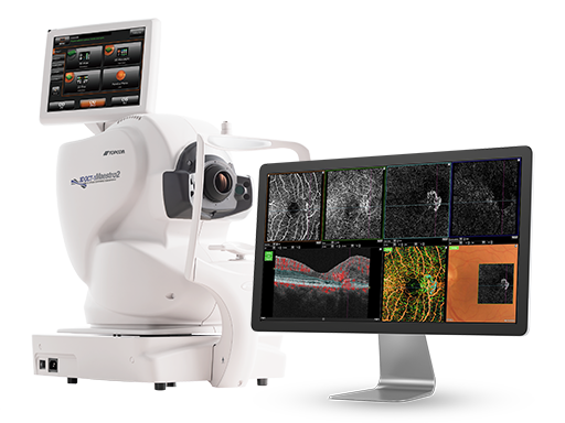
The Robotic OCT Fundus Camera with OCTA
Robotic Acquisition, Clinical Confidence
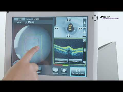
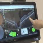
Watch this quick Maestro2 demo
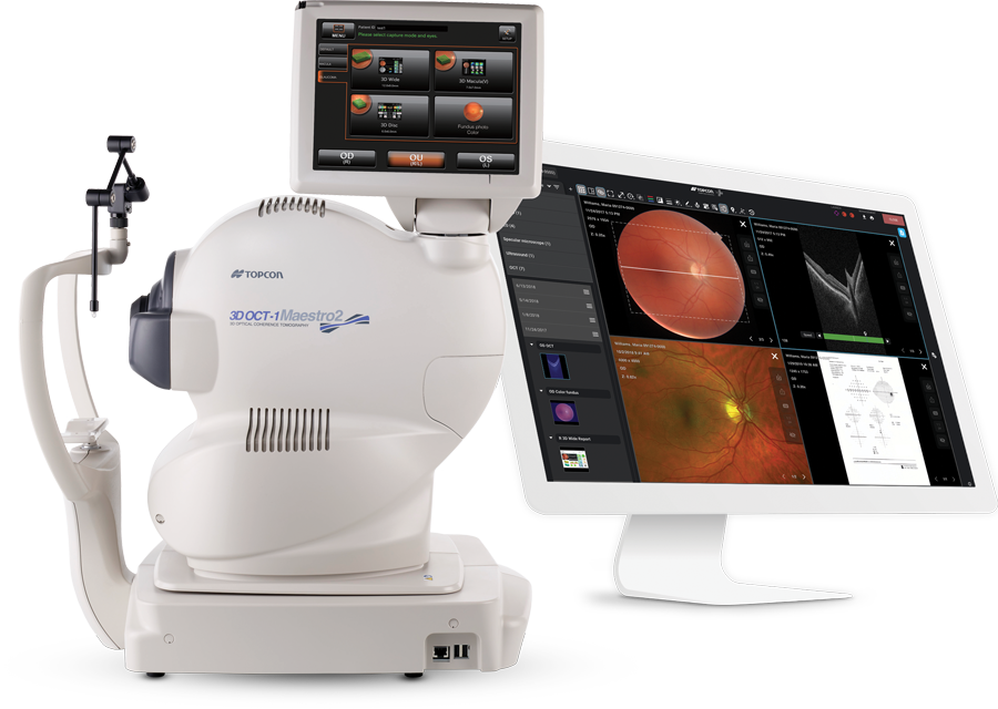
Product Features

User-friendly
Robotic OCT +
Fundus Camera

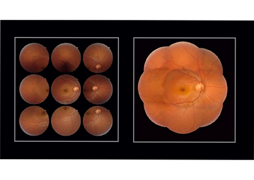
True Color¹
Fundus Photography

Full 360°
Rotating Touchscreen

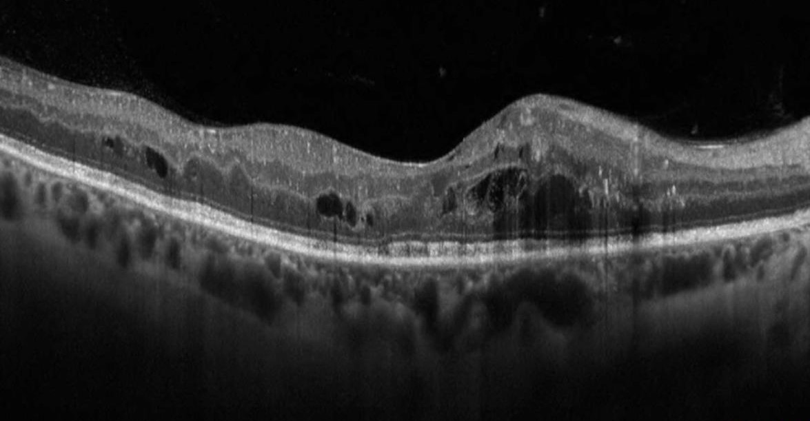
12x9mm
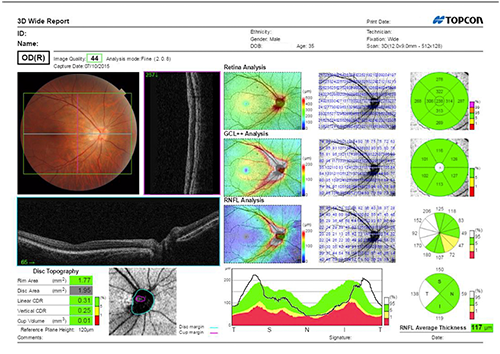
3D Wide Scan

Anterior Segment OCT

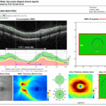
Reference Database

Single Touch Capture

OCTA²
1. True, full color fundus image simultaneously captured with white light, 24-bit color.
2. OCTA feature is optional
Topcon Maestro2 in Clinical Practice
Key Features
- USER-FRIENDLY: Robotic OCT and true color fundus camera with single-touch automated capture
- OCTA: Aerial views of the retinal vasculature are at predefined levels relevant to disease
- ANGIO B: Color-coding helps discern both normal and abnormal blood flow
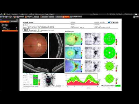
- WIDEFIELD SCAN: 12x9mm 3D wide scan captures macula and optic disc and includes the Hood Report for Glaucoma
- HIGH RESOLUTION: Multimodal Imaging with OCT and true color fundus photography*
- COMPACT FOOTPRINT: Space-saving design means the Maestro 2 Robotic OCT and Fundus Camera fits into any practice setting
*True, full color fundus image simultaneously captured with white light, 24-bit color.
Maestro2 OCT + Fundus Camera with Harmony® Clinical Data Management is a one-touch, one-screen diagnostic solution.
Ready to learn more?

Contact us today
Maestro2 OCT and Non-Mydriatic Fundus Camera Training Videos
To access our complete library of eye health education, register for Topcon Healthcare University today.


3D Wide Scan


| OBSERVATION & PHOTOGRAPHY OF THE FUNDUS | |
| Type of Photography | Color, Red-free (Note 1) & IR |
| Picture Angle for Photography | 45° ±5% or less |
| Photographable Diameter of Pupil | ø4.0mm or more : When small pupil diaphragm is not used. |
| Fundus Image Resolution (on fundus) | Center : 60 lines/mm or more |
| OBSERVATION & PHOTOGRAPHING OF THE FUNDUS TOMOGRAM | |
| Scan Range (on fundus) | Horizontal direction 3 – 12mm ±5% or less |
| OCT Scan Pattern | 3D scan (horizontal/vertical) |
| OCTA Scan Pattern | 6 x 6mm, 4.5 x 4.5mm, 3 x 3mm |
| Scan Speed | 50,000 A-Scans per second |
| Photographable Diameter of Pupil | ø2.5mm or more |
| OBSERVATION & PHOTOGRAPHING OF THE ANTERIOR SEGMENT | |
| Type of Photography | Color & IR |
| Operating Distance | 62.6 ±0.1mm (when taking a picture of anterior segment) |
| OBSERVATION & PHOTOGRAPHING OF THE ANTERIOR SEGMENT TOMOGRAM | |
| Scan Range (on cornea) | Horizontal direction 3 – 6mm ±5% or less |
| Scan Pattern | Linear scan (Line-scan/Radial-scan) |
| Scan Speed | 50,000 A-Scans per second |
Maestro2 Brochure
Reference Database Whitepaper
Maestro Clinical Compendium
OCT Reports Guide

Related Articles
Matthew Hosler, MD, Director of Glaucoma Services for the Associated…
Glaucoma, often referred to as the “silent thief of sight,”…

Watch Crystal Han, OD, of eye&I Optometry in Great Neck…
Maestro2, the robotic OCT fundus camera, now with OCTA, offers unparalleled detail in assessing both retinal structure and vascular function—all from a single scan. The Maestro2 provides OCT reference data for the optic nerve, RNFL, ganglion cell, and macular thickness. Plus, you’ll get a true-color fundus photo with pinpoint registration to the OCT.