
Empowering Providers with the Multimodal Maestro2
In 2018, Topcon Healthcare launched the Maestro2, a fully automated, compact, comprehensive system that combines spectral domain optical coherence tomography (SD-OCT) and a non-mydriatic, high-resolution retinal camera for true-color fundus photography. User uptake was rapid as clinicians recognized the broad clinical utility and benefits of the Maestro2, and it quickly became the leading product in Topcon’s OCT portfolio.
In 2019, sales of the Maestro2 topped 10,000 units, and in 2021, a new milestone was reached with 15,000 devices installed at eye care facilities around the world. Testimonials from leading ophthalmologists highlight the attributes of the Maestro2 and its value in supporting patient care.
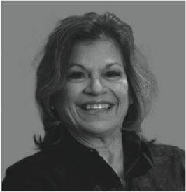
Dee Stepheson, MD, FACS, ABES, FSEE, a cataract surgeon at Stephenson Eye Associates and President of the American Board of Eye Surgery, in Venice, Florida, appreciates the efficiency of scanning with the Maestro2 and the information it provides. Dr. Stephenson said, “The Maestro2 OCT is easy to use with a short learning curve and my technicians love it! I especially like the 12x9mm wide report which includes both the macula and optic nerve along with a full color retinal photo in a single report. The Maestro2 generates beautiful, quality images that I use often for compare analysis and educating my patients. As a surgeon, I also find it to be very robust and a godsend when it comes to improving my workflow efficiency.”
Ursula Schmidt-Erfurth, MD, PhD, Head of the Department of Ophthalmology and Optometry at the Medical University of Vienna, Austria, describes the Maestro2 as an indispensable tool in her hands-on clinical practice and a reliable partner in patient care. Professor Schmidt-Erfurth said, “For a clinician coming from academia, the Maestro2 OCT is a pleasant surprise. It is easy, highly efficient and intuitive to use. It provides precise information and it is a great tool for basing treatment decisions.”

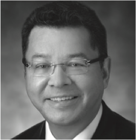
J. Fernando Arevalo, MD PhD FACS, Edmund F. and Virginia B. Ball Professor of Ophthalmology, The Johns Hopkins University School of Medicine, Baltimore, Maryland and Chairman of the Department of Ophthalmology at Johns Hopkins Bayview Medical Center, Wilmer Eye Institute. Dr Arevalo highlighted the advantage of acquiring color fundus photographs and OCT images simultaneously, “Images are easy to acquire using the Maestro2, and it provides valuable information for clinicians with a single scan.”
Recent Posts

Don’t Miss Topcon Healthcare’s Limited-Time Summer ’26 Offers
As eye care continues to evolve, practices are looking for…
Read more
Topcon Healthcare Expands Access to Standardized DICOM OCT Imaging Data
Topcon Healthcare is making its DICOM OCT Export Tool publicly available, giving its researchers a…
Read more
Powering Oculomics Research with the IDHea® Workplace Screening Dataset
Experience a new standard in ophthalmic-driven insights with the new…
Read more