
‘Must-Have’ OCT Technology for Today’s Cataract & Refractive Surgeon
As a cataract and refractive surgeon performing a high volume of surgical procedures, including minimally invasive glaucoma surgery (MIGS), I need technology that can help me keep pace with my busy practice. I’ve found that one device in particular gives me a wealth and breadth of information that is essential from a workflow and surgical planning perspective. The robotic Maestro2 OCT and Color Fundus Camera which generates a color fundus photo and OCT scan in a single acquisition, has streamlined my operations and offered important diagnostic insights.
When I am evaluating a patient post-MIGS, for example, the widefield OCT scan enables me to look at the optic nerve and macula simultaneously. This capability not only saves me time, but it offers a more complete picture of the patient’s post-op status. If the findings are normal, no other scan needs to be done at that time.
I also value the ability to take a follow-up scan, usually at about 6 weeks post-surgery, which I then compare to the initial post-op scan. This is especially useful after cataract surgery to look for any subclinical cystoid macular edema (CME) that can affect the patient’s visual acuity. Maestro2 enables me to see those kinds of subtle changes that help with my clinical decision making.
Features Helping to Increase Surgical Success
I am amazed at the things that I cannot normally see with a 90D slit lamp lens, such as epiretinal membranes (ERM) with vitreomacular traction and psuedoholes. All of these are seen so well with the Maestro2 OCT and they can be shown to the patient in 3D. This helps make me a better diagnostician and is invaluable in patient care.
For example, the device has helped me detect the presence of ERM which can have a negative effect on a patient’s cataract or refractive surgery outcome. It’s so important to know this information in advance of surgery so I can counsel the patient or send them to a retina specialist to potentially have a vitrectomy procedure first increasing the chance for better results once the patient undergoes cataract or refractive surgery.
I also appreciate Maestro2’s multi functionality and its ability to take photos of the anterior segment in patients exhibiting any evidence of pterygia, as this can induce corneal astigmatism and impact the plans for cataract surgery. The device’s anterior angle analysis software aids me in evaluating the angle pre- and post-surgery.
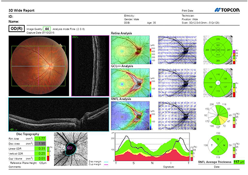
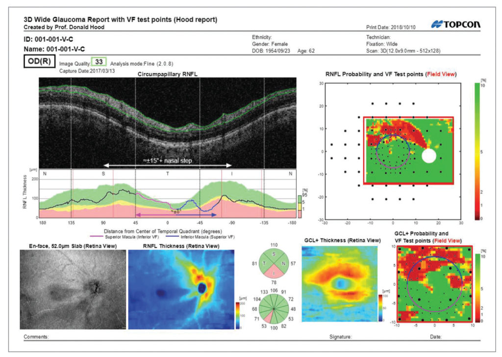
In advance of MIGS surgery, the Hood Report offers a closer look at the patient’s nerve fiber layer for better tracking of glaucomatous damage. The disc trend analysis is another tool that is extremely informative as it compares the disc parameters and retinal nerve fiber layer (RNFL) over time, enabling me to see any progression of damage. All of these capabilities help me determine which MIGS procedure is most appropriate for a given patient.
Ease of Use
Maestro2 has proven to be invaluable to my cataract and refractive surgical practice because of its ease of use. Its single-touch, robotic-capture functionality makes it a user-friendly system to learn and operate. With just one touch, the software will align, focus, and optimize the image. On occasion I need to take an OCT myself, and even I can use this device with proficiency. It helps my busy office run so much more efficiently.
The device’s built-in automation is extremely helpful, but sometimes manual or semiautomatic capture mode is needed for challenging cases or hard-to-see areas. Fortunately, this option is also easy to learn and has allowed my technicians to image patients with more complex eyes. Maestro2 is such an unbelievable machine—all of its capabilities are right at your fingertips.
Maestro2 has proven to be invaluable to my cataract and refractive surgical practice because of its ease of use. Its single-touch, robotic-capture functionality makes it a user-friendly system to learn and operate. With just one touch, the software will align, focus, and optimize the image. On occasion I need to take an OCT myself, and even I can use this device with proficiency. It helps my busy office run so much more efficiently.
Dee Stephenson, MD
An Essential Device for Patient Care
I use a pre-op OCT of the macula on every cataract patient (billing only for those with a billable diagnosis) and I am always amazed when I look at the 3D Macula Report at how much I miss with exams alone. Comparing the reports over time also gives me important information to make better decisions for patient care. I would encourage anyone considering adding another OCT to their practice to absolutely make the technology investment in the Maestro2. This compact, user-friendly machine can give you such great true-color photography and OCT with the touch of a button, is so efficient, and has such a plethora of valuable information for the clinician— from the anterior surface to the macula. Without question, this is a must-have OCT.
ABOUT THE AUTHOR
Dee Stephenson, MD, is a board-certified ophthalmic surgeon with extensive expertise in microincisional cataract surgery and implantation of premium intraocular lenses, as well as custom laser cataract techniques, and the founder of Stephenson Eye Associates in Venice, Florida.
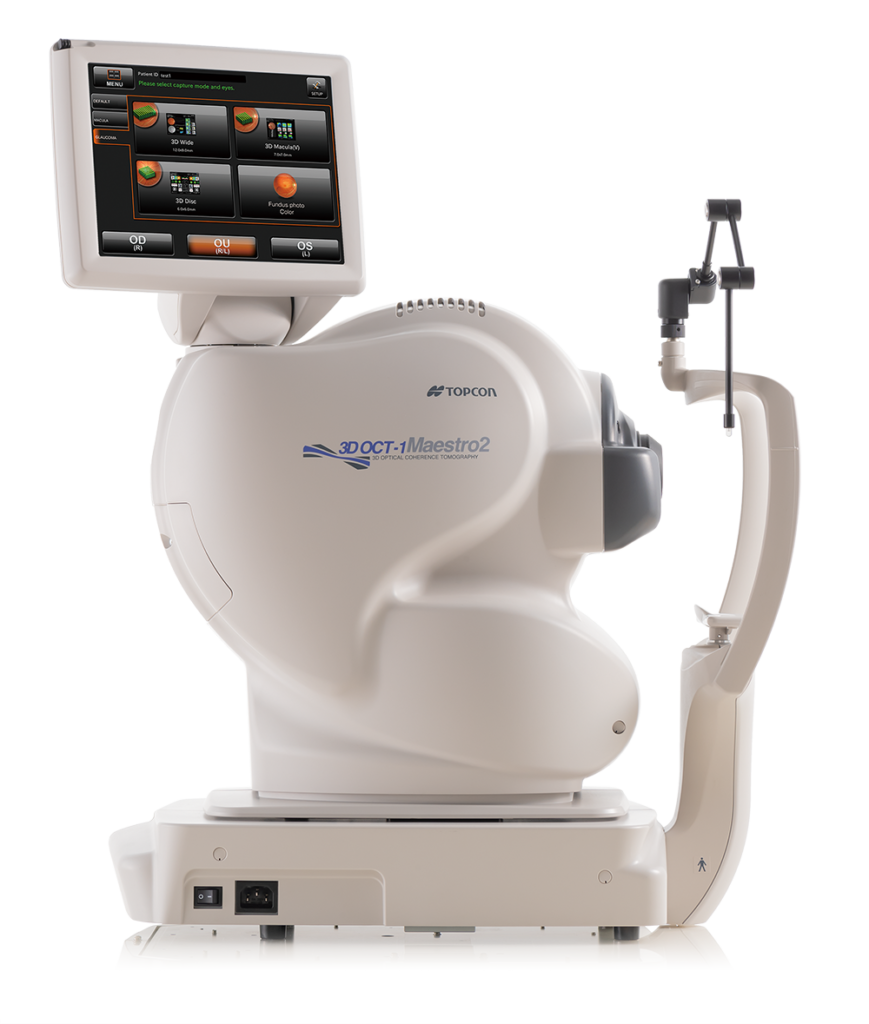
Robotic OCT +
Color Fundus Camera
Maestro2
Capture high resolution OCT scans and true color fundus images with a single touch.
Recent Posts
From Awareness to Action: Myopia Awareness Week 2025
May 19–25, 2025, marks Myopia Awareness Week, and this year’s…
Read more
The IDHea Behind Accelerating Healthcare from the Eye | Topcon Healthcare at ARVO 2025
What if we could reimagine healthcare, starting with the eye?…
Read more
Assessing Meibomian Glands for Dry Eye Diagnosis with CA-800
Maria Sampalis, OD shares why the Topcon CA‑800 Corneal Analyzer was a…
Read more