Point-of-Care OCT in the ER: How NYU Ophthalmologist Uses Topcon Maestro to Diagnose Eye Occlusions
Dr. Yasha S. Modi, Associate Professor at NYU Grossman School of Medicine, shares how the Topcon Maestro is used in their emergency room to diagnose central retinal artery occlusion (CRAO) – a time-sensitive condition requiring urgent intervention. By enabling non-ophthalmic ED technicians to capture OCT and fundus images, the Maestro allows remote retina specialists to review scans within 15-20 minutes, accelerating multidisciplinary care with neurologists and ED teams. Dr. Modi also highlights unexpected diagnoses uncovered through the system, including diabetic macular edema and exudative macular degeneration.
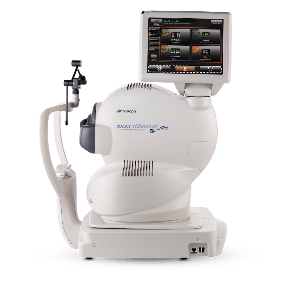
Maestro2 is the easy-to-use and affordable robotic OCT with color fundus photography.
Video Transcript
We implemented the Maestro actually for a few years ago now in our emergency room with the primary goal of actually using it to identify central retinal artery occlusion. It’s a very time-sensitive diagnosis, and the rationale for why we did that is because it allows for point-of-care access. So in other words, the retina specialist may not necessarily be in the emergency room, but if we get the images, then we can review them remotely and implement a protocol to treat patients with retinal artery occlusion.
Maestro imaging in the emergency room is performed by the ED technician. So these are technicians who are not ophthalmic trained, but because of the semi automated nature of the camera, that certainly allows for easy image acquisition. And if they need help, we have also residents in ophthalmology who are able to contribute. The primary goal of the Maestro device is the use of it to identify central retinal artery occlusion.
What’s interesting is as we use this device more and more, we’re able to identify a whole range of other diagnoses, whether it’s diabetic macular edema, central retinal vein occlusion, macular degeneration converted to the exudative form, all things that could potentially result in vision loss. The images are reviewed oftentimes within about fifteen to twenty minutes, and we look at the images and then ultimately can make a diagnosis. The nice part about it is there are hallmark features to the identification of a CRAO where we get this reflectivity in the inner retina, and that is really pathognomonic for the diagnosis.
So we know their basic vision. We then have the OCT and fundus photo features and can make a decision very quickly. The Maestro has allowed us to connect to other doctors. You know, frequently, the reason why we are oftentimes not able to treat central retinal artery occlusion in a timely fashion is because they go to the emergency room, the CT scan of the head is normal, and then they say just follow-up with your ophthalmologist in the clinic. And then unfortunately, we miss an opportunity to treat it for what it really is, which is a stroke. So adding the Maestro into the emergency room allows for a multidisciplinary approach between our ED doctors, our neurologists, and our ophthalmologists.
Recent Posts
VIDEO: How Technology is Advancing Myopia Management in Primary Eye Care
Dr. Jason Compton is dedicated to being on the front…
Read more
Advancing Glaucoma Care: A Collaborative Approach to Data-Driven Decision Support
Michael Chaglasian, OD, Associate Professor at the Illinois College of…
Read more
VIDEO: Why Axial Length Tracking is a Game-Changer for Myopia Management
Discover how Dr. Ashley Tucker elevated her myopia management practice…
Read more