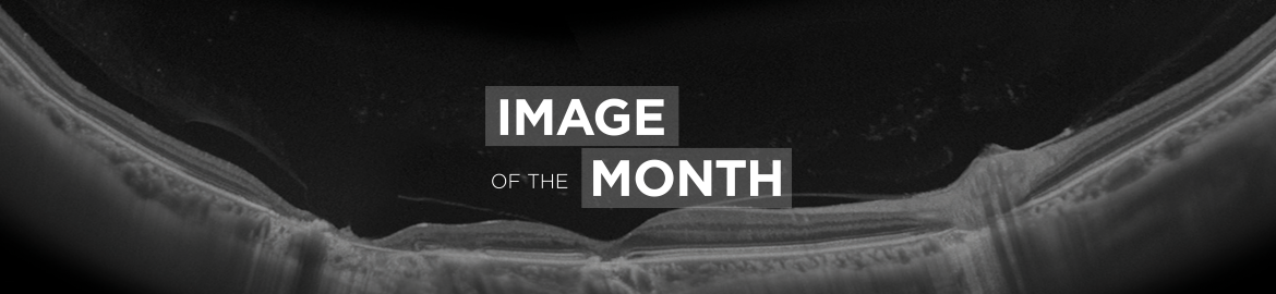
IOTM: Sub-hyaloid Hemorrhage
Image of the Month (IOTM) is a collection of interesting clinical cases with high quality images for all relevant imaging modalities (ex: color fundus, OCT, OCTA, FAF, FA, En Face, Red-free, choroidal vasculography (CVG), anterior imaging) and other clinical results if relevant (ex: visual field plots). Each case is submitted by an eye care professional using one of Topcon’s industry-leading OCT devices.
Case background
This case shows the benefits of using Swept Source OCT technology when imaging through media opacities such as sub-hyaloid hemorrhage. Using the Topcon DRI Swept Source OCT’s 1050nm wavelength it is possible to image all the retinal layers of a 64-year-old male patient, as well as the choroidal structures through the hemorrhage while maintaining a clear imaging of the vitreal structures such as the posterior vitreous detachment and the hemorrhage beneath it. The ability to clearly image the choroid, even in cases with media opacities, is becoming increasingly important. The upper images (A and B) show a fundus image (A) as well as a horizontal OCT (B) scan through a sub-hyaloid hemorrhage under a posterior vitreous detachment as well as through a much denser pre-retinal hemorrhage. The bottom images (C and D) show the same case using a vertical OCT scan.
—Carl Glittenberg, MD, FEBO
Diagnosis: Proliferative Diabetic Retinopathy with Sub-Hyaloid Hemorrhage
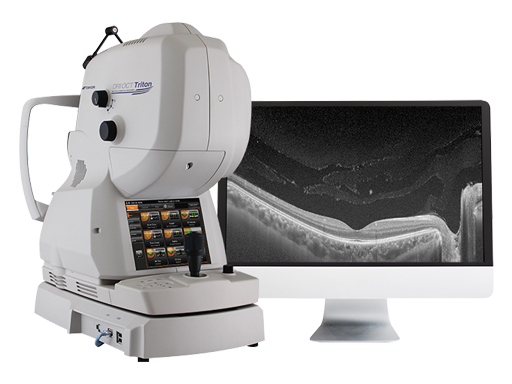
Captured with: Topcon DRI OCT Triton
DRI OCT Triton Images:
A–B. Color Fundus and Horizontal SS-OCT Scan
C–D. Color Fundus and Vertical SS-OCT Scan


Increase diagnostic capabilities with the multi-modal swept source OCT
The opinions, ideas, views and assumptions expressed are the author’s own and do not necessarily represent the views of Topcon, nor do they constitute advice from Topcon.
Recent Posts

IOTM: Choroidal Nevus with Drusen
This 52 year-old patient presented with a sudden central visual…
Read more
IOTM: Branch Retinal Artery Occlusion
This 52 year-old patient presented with a sudden central visual…
Read more
IOTM: Toxoplasmosis Retinochoroiditis
A 57-year-old Indian female with a prior history of posterior…
Read more